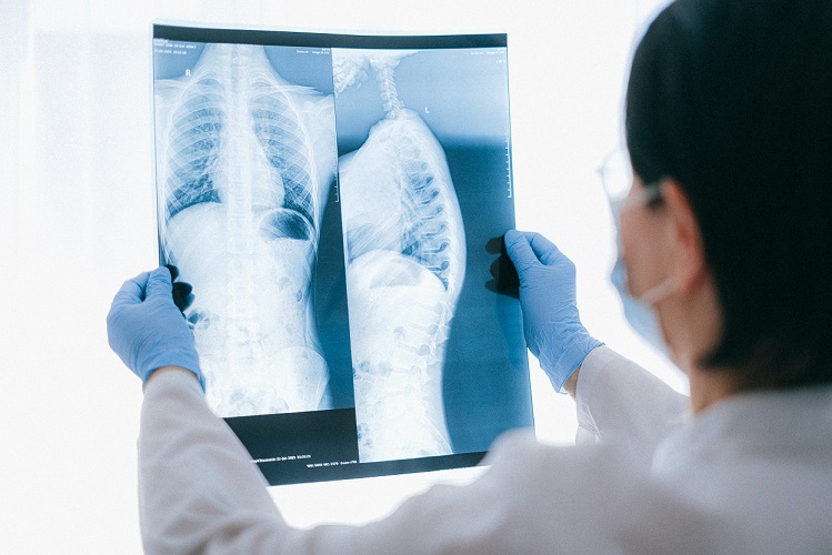
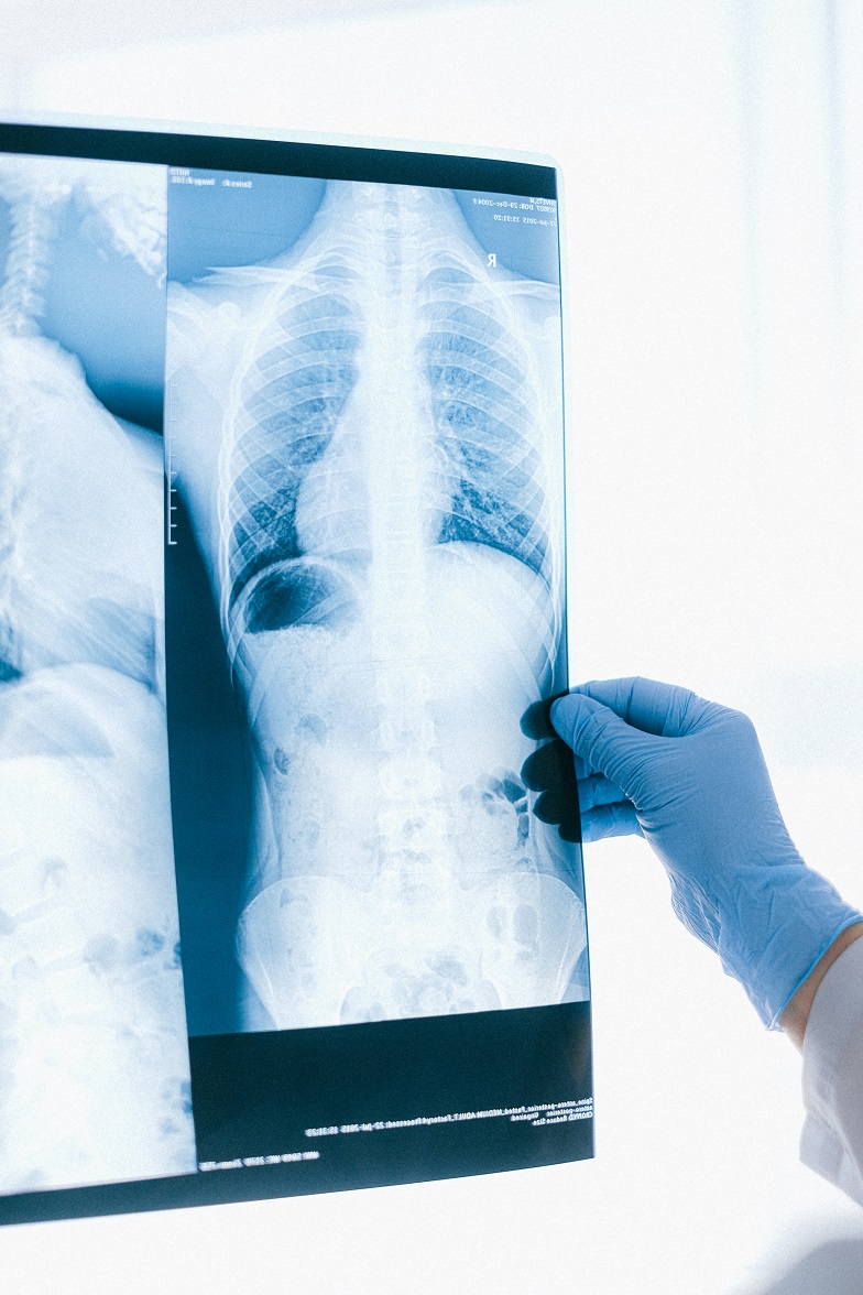
Chest scans are able to help determine how long lung cancer patients will live.
According to a recent report published online in the American Journal of Roentgenology, CT chest scanning can predict the long-term prognoses of patients with Stage 1 lung cancer “better than clinical characteristics” alone. In Stage 1, a malignant tumor is present in a single lung (rather than both lungs) and has not yet traveled to a person’s lymph nodes or other internal organs. Historically, experts have surmised that patients have a five-year survival rate after their initial diagnosis. Of course, catching the cancer at a later stage could decrease this period of time significantly.
“Survival rates are determined by biological age, Eastern Cooperative Oncology Group (ECOG) score, and the presence of comorbidities,” Senior study author Florian J. Fintelmann, MD, radiologist at Massachusetts General Hospital and associate professor of radiology at Harvard Medical School, Boston, Massachusetts, explained.

The retrospective study analyzed the CT chest scanning results from 282 patients (168 of whom were female and 114 were male, with median age of 75 years) with Stage 1 who were treated with SBRT in the eight-year span between January 2009 and June 2017. The authors cite, “Associations of clinical and imaging features with OS were quantified using a multivariable Cox proportional hazards (PH) model. Penalized multivariable Cox PH models to predict OS were constructed using clinical features only and using both clinical and imaging features. Models’ discriminatory ability was assessed by constructing time-varying ROC curves and computing AUC at prespecified times.”
Furthermore, they wrote, “Modeling that incorporates noncancerous imaging features captured on chest computed tomography (CT) along with clinical features, when calculated before stereotactic body radiation therapy (SBRT) is administered, improves survival prediction compared with modeling that relies only on clinical features.”
Fintelmann said, “The focus of the study was to look at the environment in which the cancer lives. This is looking at parameters like the aortic diameter, body composition – that is, the quantification and characterization of adipose tissue and muscle – coronary artery calcifications, and emphysema quantification. CT images are used by radiation oncologists to determine where the radiation should be delivered. There is more information from these images that we can utilize.”
Pre-treatment chest images with CT specifically allowed the investigative team to assess coronary artery calcium (CAC) score, pulmonary artery (PA)-to-aorta ratio, emphysema, and several measures of body composition. The team put together a statistical model to show the correlation between clinical and imaging features and overall patient survival.
“An elevated CAC score (11-399: HR, 1.83 [95% CI, 1.15 – 2.91]; ≥ 400: HR, 1.63 [95% CI, 1.01 – 2.63]), increased PA-to-aorta ratio (HR, 1.33 [95% CI, 1.16 – 1.52], per 0.1-unit increase) and decreased thoracic skeletal muscle (HR, 0.88 [95% CI, 0.79 – 0.98], per 10 cm2/m2 increase) were independently associated with shorter overall survival,” they found. The team also reported that the five-year survival model was “superior for the model that included clinical and imaging features and inferior for the model restricted to only clinical features.” Overall, CT chest scanning was reliable in predicting survival timeframes of those with Stage 1.
Sources:
What are the stages of lung cancer?
Clinical Chest Images Power Up Survival Prediction in Lung Cancer


Join the conversation!