New method helps map skin cancer more clearly before surgery.
A team of scientists in Singapore may have found a new way to help doctors see skin cancer more clearly and treat it more effectively. This approach uses a special kind of imaging that can create clear, three-dimensional pictures of tumors inside the skin. The method combines light and sound to build a detailed image of what’s happening beneath the surface. It helps doctors see the true shape and depth of the tumor without needing to cut the skin open first. They tested it on patients with the most common kind of skin cancer, called basal cell carcinoma, or BCC for short.
Basal cell carcinoma is the kind of skin cancer that many people get, especially as they get older. In Singapore, cases have been going up, and older adults are seeing it more often. Usually, when someone has a suspicious spot on their skin, they need a biopsy to know if it’s cancer. That means a small piece of skin is removed and checked under a microscope. If it’s cancer, the doctor might remove the tumor using a method called Mohs surgery, where they cut away the skin little by little and check each piece under a microscope until no more cancer is found. While this is effective, it can take a long time and be uncomfortable. Some people even need more than one surgery if the full tumor wasn’t removed the first time.
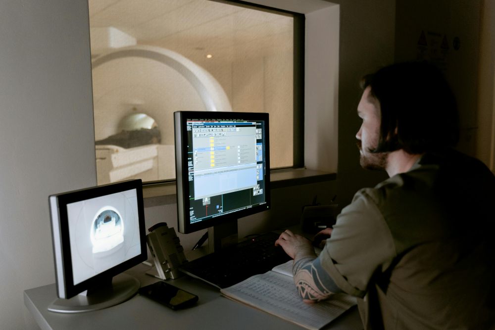
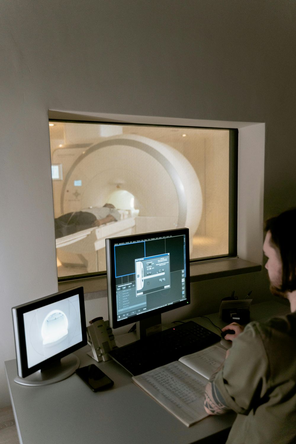
The new technique hopes to change all that. Scientists from Singapore’s Agency for Science, Technology and Research, along with doctors from the National Skin Centre, have worked together to test a new machine that uses something called multispectral optoacoustic tomography. That’s a mouthful, but the idea is simple. The machine sends light into the skin, which causes a tiny bit of heat. This heat creates sound waves that bounce back and help build a picture. Then, an automated computer program outlines the tumor’s shape. The system shows how wide, deep, and tall the tumor is—like building a 3D map of it inside the body. This helps the doctor see exactly how much needs to be removed, which makes surgery faster and more precise.
In the first test of this technology, eight patients had their tumors scanned before surgery. The scans matched up closely with the results doctors got after surgery using regular methods. That’s a good sign that the machine is doing its job well. It also showed that the system could help avoid extra surgeries by getting things right the first time. Fewer surgeries mean less pain and shorter recovery for patients.
While this new imaging tool was made for basal cell carcinoma, the Singapore researchers believe it could help with other types of skin cancer too. The team is still testing it, but they’re hopeful that this could be the start of something bigger. If the system keeps performing well, doctors may soon have a new way to treat skin cancer that’s faster, easier, and more accurate.
For now, the team will keep testing and improving the system. They want to make sure it works well for different skin types and in all kinds of cases. But if everything goes well, this could become part of regular cancer care, giving patients a smoother path to treatment and recovery.
Sources:
-New 3D imaging technique enhances basal cell carcinoma diagnosis
Join the conversation!